

This article is based on the scientific preprint “AI Skin Analysis in Real-World Practice: A Multi-Source Evidence Assessment” by Viktor Shpudeiko and Kirill Sokolov (Skinive BV), published on Research Square. DOI: 10.21203/rs.3.rs-11727541/v1. Not yet peer-reviewed.
Abstract
Relevance. AI is increasingly used to assess skin lesions, but trust in it depends not only on laboratory accuracy but also on transparent data about real-world performance. Most studies show high accuracy under controlled conditions, but few combine technical validation, independent clinical evaluation, and comparison with scientific literature in a single report.
Objective. To evaluate the capabilities and limitations of AI skin analysis based on three sources: the Skinive Accuracy Report 2026, an independent clinical evaluation at a specialized referral center, and current peer-reviewed publications.
Methods. Synthesis of data from technical validation of the Skinive algorithm, independent clinical evaluation on a complex patient sample, and analysis of scientific literature.
Results. The Skinive algorithm shows progressive improvement in accuracy from version to version. In the independent evaluation on a complex sample, a clinically useful result (correct diagnosis in the top 3, correct disease group, or correct referral direction) was achieved in 80% of cases.
Conclusions. AI skin analysis can be clinically meaningful support in the early assessment of lesions, but as a decision-support tool, not a replacement for a physician. Further prospective multicenter studies are needed.
Introduction
AI has become one of the fastest-growing technologies in dermatology. Deep learning makes it possible to achieve accuracy close to that of experienced dermatologists under controlled conditions. However, translating these results into routine practice remains a difficult task.
Most studies evaluate AI using standard metrics (sensitivity, specificity, AUROC), but these do not reflect how the system performs with different image quality, rare diseases, and diverse patients. Therefore, increasing attention is being paid to Real-World Evidence (RWE) — data from real-world practice.
This report combines technical validation, independent clinical evaluation, and comparison with the literature. It does not claim that AI makes a final diagnosis, but rather considers it as a decision-support tool: early risk assessment, self-screening, and prioritization of referrals to a specialist.
Sources of Evidence
The report combines three independent sources:
- Skinive Accuracy Report 2026 — technical validation of the algorithm using a standardized methodology.
- Independent clinical evaluation at a specialized referral center on a complex patient sample.
- Current scientific literature on AI in dermatology and digital health.
Each source provides different information: technical validation — performance under standard conditions; clinical evaluation — behavior in complex cases; literature — external context.
Real-World Insights from 6.5 Million AI Skin Checks
Understanding How AI Is Used in Everyday Dermatology
Since launch, the Skinive platform has performed more than 6.57 million AI skin analyses (Q3 2026) — one of the largest proprietary collections of real-world smartphone-based skin assessments. These data do not reflect disease prevalence, but they show how people use AI dermatology in real life.
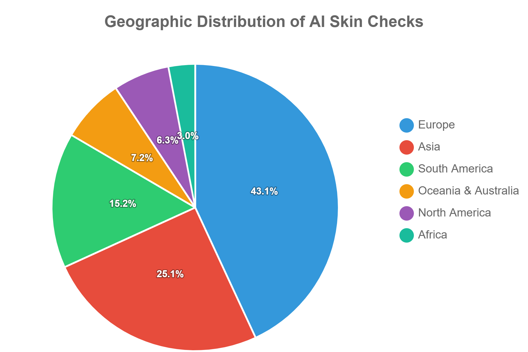
Figure: Geographic Distribution of AI Skin Checks
Key interpretation: The dataset represents routine use across multiple healthcare systems and geographic regions, increasing the diversity of real-world operating conditions.
Geography. Most analyses come from Germany, Russia, India, the United Kingdom, France, Italy, Bangladesh, Spain, Australia, and Brazil. Next are Argentina, Colombia, the Netherlands, Mexico, Poland, Belarus, the United States, Austria, Chile, and the Philippines.
What Users Actually Check
Users use AI not only for skin cancer screening. Among the findings identified, inflammatory skin diseases accounted for the largest share.
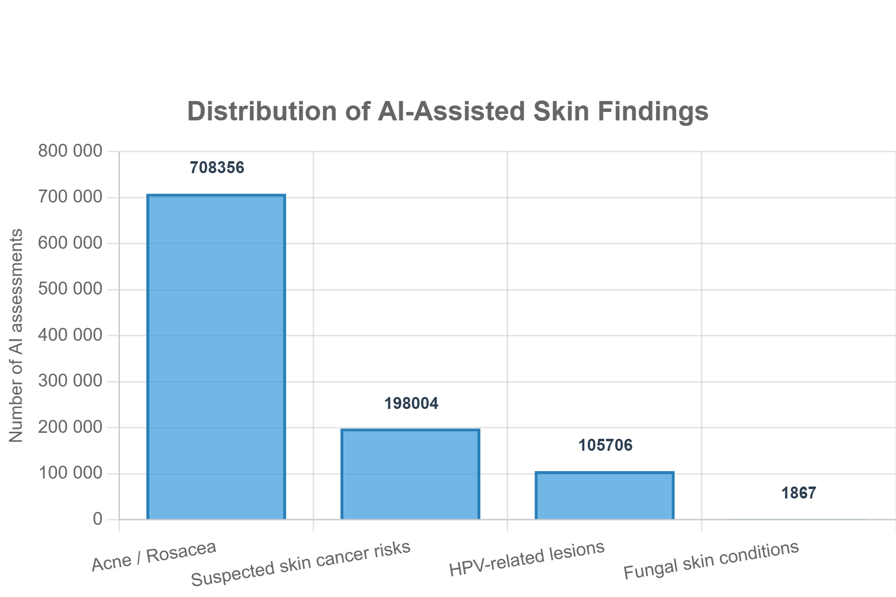
Figure: Distribution of AI-Assisted Skin Findings
Key interpretation: Inflammatory skin conditions constitute the largest category of real-world AI usage, while skin cancer risk assessment remains a major clinical application.
Clinically Relevant Risk Detection
In 14.68% of all AI analyses, findings were identified that were classified as clinically significant risk indicators requiring additional attention. These are not confirmed diagnoses, but the algorithm’s recommendations about the possible need to consult a specialist.
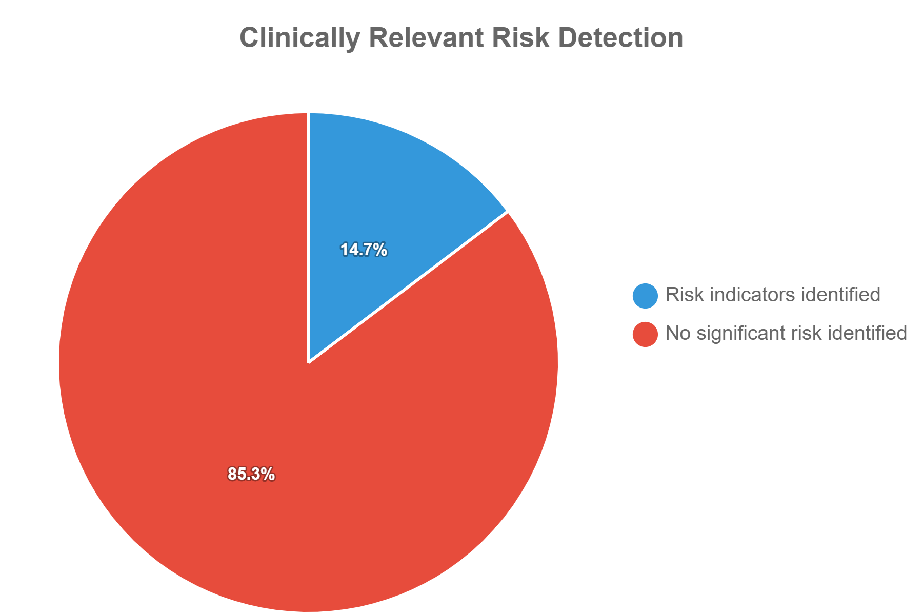
Figure: Clinically Relevant Risk Detection
Key interpretation: Proportion of completed AI skin assessments resulting in clinically relevant risk recommendations. Higher risk detection rates likely reflect self-selection of users choosing to evaluate suspicious skin findings.
Mobile Platforms and Accessibility
About 60–65% of analyses were performed on Android, the rest on iPhone. This indicates the broad accessibility of smartphone dermatology across different ecosystems.
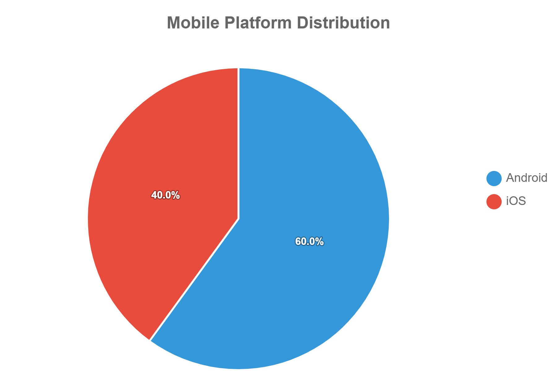
Figure: Mobile Platform Distribution
Image Quality Remains the Largest Modifiable Factor
Among failed analyses, most are associated with two preventable factors:
- insufficient lighting;
- image blur (poor focus or camera movement).
Unlike limitations related to lesion morphology or the camera, users can correct these factors themselves.
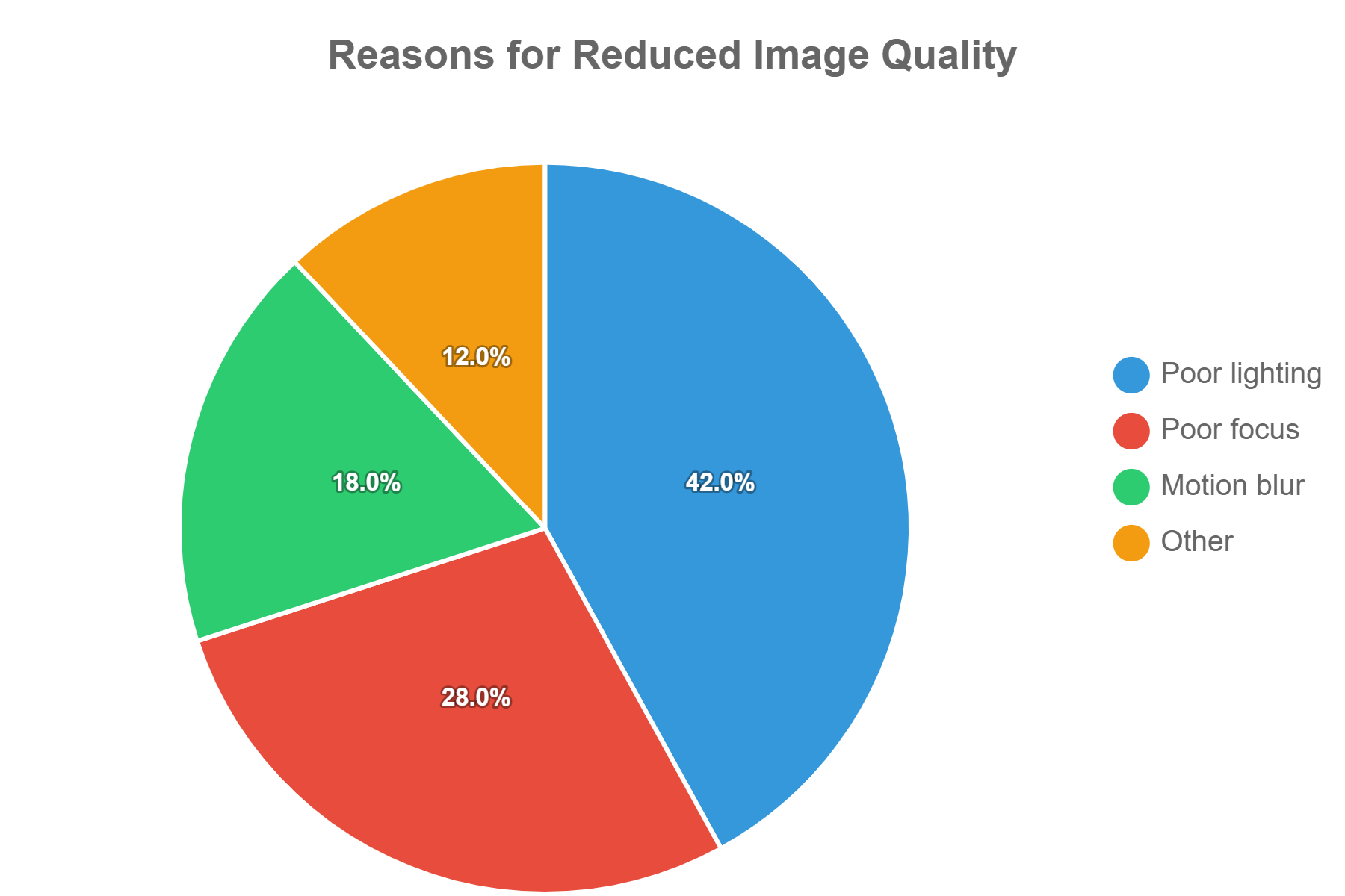
Figure: Reasons for Reduced Image Quality
Key interpretation: Educational guidance on image acquisition may further improve diagnostic performance without requiring changes to the AI algorithm itself.
Key Real-World Observations
Analysis of more than 6.57 million AI-assisted skin assessments provides several important observations that extend beyond conventional validation studies.
- AI dermatology is used more often for inflammatory diseases than many clinicians expect.
- Users turn to AI for specific concerns, not for mass screening.
- Image quality is the main modifiable factor for successful analysis.
- The accumulation of proprietary clinical data made it possible to continuously improve the algorithm, especially in recognizing pigmented lesions and skin cancer risk.
Accuracy Report 2026: Key Findings
The report documents the evolution of the Skinive algorithm using a standardized methodology applied consistently from version to version.
- The algorithm recognizes 55 skin diseases.
- Training was performed on a clinically annotated dataset of about 250,000 images.
- The platform has processed more than 6.5 million AI analyses since launch.
- A consistent increase in sensitivity, specificity, accuracy, and F1-score is noted.
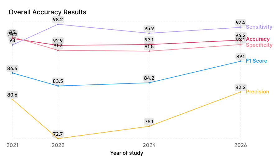
Figure: Evolution of Skinive AI performance (2021–2026)
Key interpretation: Improvements were achieved without optimizing one metric at the expense of others — the profile has become more balanced. This is critical for decision-support systems, where a balance between missed lesions and false positives matters.
Clinical Stress-Test in a Specialized Referral Center
Study Design
In 2026, Skinive underwent clinical evaluation at a specialized referral center (the name is not disclosed due to confidentiality terms). The center receives complex cases from general practitioners and dermatologists. This is a stress test, not population screening.
- A total of 241 examinations were performed.
- Complete data are available for 160 cases (the main analytical cohort).
- Reference diagnosis: histopathology — 117 cases, cytology — 22, dermatologist’s clinical diagnosis — 21.
Evaluation was conducted at several levels:
- Exact match with top-1, top-2, top-3;
- Correct disease group;
- Correct referral direction by risk;
- Complete mismatch;
- Cases of diseases not covered by the algorithm.
Differences between basal cell and squamous cell carcinoma, as well as between melanoma and lentigo maligna melanoma, were not considered errors — they are difficult to distinguish visually and require histology.
Patient Population
The sample was extremely selective and complex: suspected melanoma, basal cell and squamous cell carcinoma, Bowen’s disease, and dysplastic nevi predominated. Simple benign lesions were virtually absent.
This is important for interpretation: in ordinary screening populations, most lesions are benign and typical. Here, however, there was a disproportionate number of atypical, malignant, precancerous, and diagnostically ambiguous cases requiring differential diagnosis. Therefore, the results cannot be directly extrapolated to mass screening.
Results
- Exact top-1 match: 87 of 160 cases — 54.4%.
- Exact top-3 match: an additional 19 cases (11.9%) matched in second position and 7 (4.4%) in third. In total, the correct diagnosis was in the top 3 in 113 of 160 cases — 70.6%.
- Correct disease group: another 6 cases (3.75%) — the exact diagnosis did not match, but the disease group was correctly identified.
- Correct referral direction: 9 cases (5.6%) — the diagnosis and group did not match, but the risk assessment led to the correct referral decision.
- Clinically useful composite result: when combining top-3, correct group, and correct direction, Skinive produced a clinically useful result in 128 of 160 cases — 80.0%.
- Remaining cases: 6 unrecognized diseases (3.75%) and 26 completely incorrect results (16.25%).
- Subgroup analysis: in 23 cases without histology or with indeterminate cytology, the top-3 matched the dermatologist’s clinical diagnosis in 19 cases (82.6%), with discrepancies in 4 (17.4%), three of which were diseases not recognized by the algorithm.
Clinical Interpretation
The main conclusion is not that Skinive made an exact diagnosis in every case — it did not. The key point is that the system retained useful diagnostic or triage information in 80% of an unusually complex referral sample.
For a decision-support tool, exact match and correct referral direction are related but different outcomes. Histopathology remains necessary for the final classification of many malignant and borderline lesions. An AI response based on a photograph of skin pathology should not be evaluated solely by how accurately it reproduces a histological label. Its ability to raise suspicion, assign risk, and prompt timely specialist examination is also clinically significant.
Error profile: completely incorrect results — 16.25%, mainly rare conditions (Spitz nevus, juvenile xanthogranuloma, pyogenic granuloma) or atypical manifestations of common diseases. There were more false-positive results than false-negative ones.
A tendency toward false-positive results is preferable from the standpoint of oncological vigilance — the risk of ignoring a dangerous lesion is reduced. But false positives are not harmless: anxiety, unnecessary consultations, and additional burden on the system. The balance depends on clinical conditions and the availability of specialists.
The results support the use of Skinive as an auxiliary tool for risk awareness and decision support, rather than as an autonomous diagnostic system.
Study Limitations
- The main cohort is only 160 cases, which limits statistical precision.
- The sample is not consecutive or population-based, and is enriched with complex and malignant lesions — spectrum and selection bias.
- Not all reference diagnoses were histologically confirmed.
- The composite 80% combines three different levels; it cannot be called “80% diagnostic accuracy.” Strict top-1 and top-3 are 54.4% and 70.6%.
- The results cannot be directly transferred to mass screening, primary care, or independent user self-assessment.
Comparison with Published Literature
Over the past decade, many studies have shown that deep learning reaches dermatologist-level performance in individual classification tasks under controlled conditions. But these same works emphasize that laboratory accuracy does not equal effectiveness in routine practice.
Systematic reviews note high heterogeneity across studies: differences in image acquisition, disease prevalence, reference standards, and metrics make direct comparison difficult. Therefore, prospective clinical validation and RWE are recommended.
The Skinive results are consistent with this trend: gradual improvement through iterative development, dataset expansion, and continuous validation, rather than a single technological breakthrough. The report does not attempt to prove superiority over other systems — differences in design and samples preclude valid comparison. The conclusions support the consensus: reliable evaluation of clinical decision-support systems requires several complementary sources of evidence.
Discussion
The report combines three sources: technical validation, independent clinical evaluation, and comparison with the literature.
Technical validation shows progressive improvement of the algorithm. Clinical evaluation extends these data by showing behavior under substantially more complex conditions. The literature provides external context.
An important observation: evaluation of medical AI must go beyond a single metric. Exact match remains important, but it does not fully reflect the clinical value of systems for early assessment and triage. Even in a complex referral population, Skinive often provided clinically meaningful support through differential diagnosis, group recognition, or referral recommendation.
Practical implications for primary care, teledermatology, and self-assessment: the main question is often not “what is the exact diagnosis,” but “does the patient need to be referred to a physician.” When used properly, AI can support early detection, awareness, and prioritization of referrals, complementing rather than replacing examination, dermatoscopy, and histology.
The report emphasizes the importance of transparency: technical accuracy alone provides an incomplete picture. Independent evaluation, discussion of limitations, and comparison with the literature allow realistic interpretation of the results.
Limitations and Future Directions
- The clinical evaluation was conducted at a specialized center in an extremely selective population — the results do not reflect routine primary care or self-screening.
- Not all lesions underwent the same diagnostic verification.
- The closed nature of the full dataset limits external reproducibility, although the methodology and metrics are disclosed.
- No single dataset, study, or metric can fully characterize the reliability of an AI system. Further RWE, independent external evaluations, and prospective multicenter studies are needed.
- Future versions of the report plan to include larger prospective datasets, longitudinal analysis, additional external evaluations, and expanded data on patient triage.
Conclusion
AI is becoming an important part of modern dermatology, but trust in it depends on transparent evidence, not isolated metrics.
Main conclusions:
- Technical validation demonstrates continuous improvement of the Skinive algorithm from version to version.
- Independent clinical evaluation confirms clinically meaningful benefit in complex conditions: 80% clinically useful results in an unusually complex sample.
- Clinical usefulness goes beyond exact diagnosis: correct classification and referral recommendations are important components of decision support.
- The results are consistent with the literature, which increasingly emphasizes the need to combine technical validation with RWE and independent clinical evaluation.
- Transparency about strengths and weaknesses is essential. Responsible implementation of AI requires continuous validation, independent evaluation, and regular reporting.
AI does not replace a dermatologist, but supports early risk assessment, patient awareness, and prioritization of referrals when integrated into existing standards of care.
Data Availability Statement
The full training dataset and proprietary image collections are not publicly available due to protected clinical data and intellectual property. Aggregated metrics, methodology, and validation results are available via the Skinive Accuracy Report 2026 and scientific publications.
Conflict of Interest
Kirill Sokol is the founder of Skinive. The authors are involved in the development, validation and commercialization of the Skinive AI platform.
The independent clinical assessment described in this report was conducted by clinicians at a specialized tertiary referral dermatology center. The medical institution retained full responsibility for establishing reference diagnoses according to routine clinical practice. The identity of the institution is withheld under a confidentiality agreement and has no influence on the interpretation of the reported findings.
The authors declare no other competing interests.
Frequently Asked Questions
What is the Skinive Real-World Evidence Report?
The Skinive Real-World Evidence Report summarizes findings from more than 6.5 million AI-assisted skin checks, combined with clinical validation data and peer-reviewed scientific evidence. Its purpose is to transparently describe how Skinive AI performs in everyday use, its diagnostic accuracy, and the limitations of large real-world datasets.
Why is a dataset of 6.5 million skin checks important?
Large real-world datasets expose AI algorithms to a broad range of skin types, imaging conditions and clinical presentations that are difficult to reproduce in controlled studies. They also provide valuable insights into how people actually use AI-assisted dermatology in routine practice.
Can AI accurately detect skin cancer?
Artificial intelligence can help identify skin lesions that may require medical attention, including suspicious pigmented lesions associated with melanoma and other skin cancers. However, AI should be considered a decision-support tool rather than a replacement for clinical examination or histopathological diagnosis.
Can AI replace a dermatologist?
No. AI is designed to assist with early risk assessment and triage, not to replace dermatologists. Final diagnosis and treatment decisions should always be made by qualified healthcare professionals.
What factors most commonly reduce AI performance in skin analysis?
The most common causes of unsuccessful or lower-quality image analysis are:
- poor lighting,
- image blur,
- incorrect focus,
- incomplete visualization of the lesion.
Improving image quality significantly increases the reliability of AI-assisted assessment.
Is Skinive clinically validated?
Yes. Skinive has published multiple validation studies and an annual Accuracy Report describing the methodology, validation datasets and diagnostic performance of the algorithm. Clinical evidence is continuously updated as theplatform evolves. For more information please visit Clinical Evidence Hub
Which smartphones were used for these AI skin checks?
Approximately 60–65% of examinations were performed on Android devices, while the remaining assessments originated from iPhone users. This broad device distribution helps evaluate algorithm performance across different camera systems and real-world imaging conditions.


