

A new spot, bump or patch on your skin can raise questions. Is it a mole, a skin tag, a cyst or something else? Many common skin lesions are benign, but different conditions can look similar, and appearance alone does not always reveal what a lesion is.
This guide explains how some common skin growths and spots may look, what changes to watch for and when to seek medical advice. You can also explore related information and pictures in the Skinive Dermatology Atlas.
If a spot is new, changing, bleeding or otherwise concerning, have it assessed by a qualified healthcare professional.
What Is a Skin Lesion?
A skin lesion is an area of skin that looks or feels different from the surrounding skin. The term covers many kinds of changes, from a flat pigmented spot to a raised bump or a lump beneath the skin.
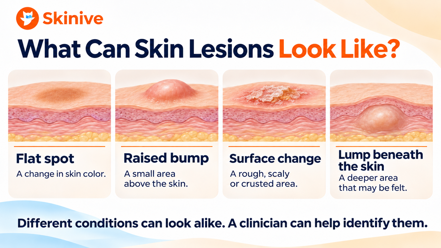
A lesion may be:
- Flat, like a freckle or sunspot
- Raised, like a skin tag or certain moles
- Rough or scaly, like some types of keratosis
- Filled with material, like a cyst
- Located beneath the skin, like a lipoma
Skin lesions can vary in color, shape, size and texture. Their appearance may also differ across skin tones.
The word lesion does not mean cancer. It is a general medical term for a change in the skin.
What Do Common Skin Lesions Look Like?
Many benign skin lesions have recognizable features. However, a description or picture cannot confirm a diagnosis. Some benign growths resemble skin cancer, while a concerning lesion may not look like the examples you have seen online.
The sections below describe common appearances and explain when professional assessment is useful.
Seborrheic Keratosis
Seborrheic keratosis is a common benign skin growth that often appears in adulthood.
It may look:
- Light tan, brown, dark brown or close to the surrounding skin color
- Slightly raised, with a waxy, rough or wart-like surface
- As though it is “stuck on” the skin
- Like a single growth or one of several similar spots
Seborrheic keratoses can occur on the face, neck, chest, back and other areas. A confirmed seborrheic keratosis usually does not need treatment unless it becomes irritated or a person wants it removed.
When to seek advice: Have a new, rapidly changing, bleeding or unusual growth examined. Skin cancer and other conditions can sometimes resemble a seborrheic keratosis.
Dermatosis Papulosa Nigra
Dermatosis papulosa nigra causes small, often multiple, dark brown or black raised spots. They commonly appear on the cheeks, around the eyes or elsewhere on the face and neck.
These benign growths are more frequently seen in people with darker skin tones. They may begin during adolescence or early adulthood and become more numerous over time.
They usually do not require treatment. If removal is desired, consult a dermatologist, as procedures can sometimes leave changes in skin color or scars.
Dermatosis papulosa nigra is related to seborrheic keratosis, although its appearance can differ.
Stucco Keratosis
Stucco keratoses are small, rough, whitish or grayish bumps that often appear on the lower legs, ankles or tops of the feet. They may look as if they are sitting on the surface of the skin.
These growths are generally benign and often cause no symptoms. Because other scaly lesions can look similar, ask a healthcare professional to examine a new, changing or uncertain spot.
You can explore related benign skin growths in the Skinive Dermatology Atlas.
Skin Tags
A skin tag, also called an acrochordon, is a small, soft growth that may hang from the skin on a narrow stalk. Skin tags can be skin-colored or darker and commonly appear in areas where skin folds or clothing rubs, such as the neck, armpits, eyelids or groin.
A skin tag may become irritated by jewelry, clothing or shaving. Once a healthcare professional has confirmed what it is, removal can be considered if it causes discomfort or for cosmetic reasons.
Do not cut, burn or remove a skin tag yourself. A growth that looks like a skin tag may be something else, and home removal can cause bleeding, infection or scarring.
Cherry Angiomas
Cherry angiomas are small benign growths made up of blood vessels. They often look like red, reddish-purple or darker bumps and can be flat or slightly raised.
They commonly appear on the trunk and may become more numerous with age. A confirmed cherry angioma usually does not need treatment, although a clinician can discuss removal if it is repeatedly irritated or cosmetically bothersome.
A new, changing or bleeding red or dark spot should be examined rather than assumed to be an angioma.
For related information about vascular skin growths, see cutaneous hemangiomas in the Skinive Dermatology Atlas.
Dermatofibroma
A dermatofibroma is a benign, firm skin growth. It often appears on the legs, although it can occur elsewhere.
A dermatofibroma may be:
- A small, firm bump
- Brown, reddish-brown, pink or purple, depending on the skin and the lesion
- Slightly raised or relatively flat
- Sometimes indented when gently pinched from the sides during a clinical examination
The appearance can vary. A new, growing, painful or otherwise unusual lump should be assessed by a healthcare professional.
Solar Lentigo: Sunspots and Age Spots
A solar lentigo, often called a sunspot or age spot, is a flat pigmented patch associated with sun exposure. It can range from light brown to darker brown and often appears on the face, hands, shoulders or other sun-exposed areas.
A confirmed solar lentigo is benign. However, it can be difficult to distinguish a sunspot from another pigmented lesion without an examination. A spot that is new, changing or different from others should be checked.
Learn more about lentigo in the Skinive Dermatology Atlas.
Sunspots can reflect previous UV exposure. Protecting your skin from further excessive UV exposure remains important, whether or not you have visible sunspots.
Sebaceous Hyperplasia
Sebaceous hyperplasia occurs when an oil gland becomes enlarged. It often appears as a small, skin-colored or yellowish bump, sometimes with a slight indentation in the center.
These bumps commonly occur on the forehead or central face. Sebaceous hyperplasia is benign, but some other skin lesions, including basal cell carcinoma, can look similar.
A dermatologist can examine an uncertain bump and advise whether any further testing is needed. Treatment is not always necessary after the diagnosis is confirmed.
Epidermoid Cysts
An epidermoid cyst, sometimes called an epidermal inclusion cyst, is a lump beneath the skin containing keratin. People sometimes call it a “sebaceous cyst,” although that name is not always accurate.
An epidermoid cyst may:
- Feel like a round lump beneath the skin
- Grow slowly
- Have a small central opening
- Become tender, inflamed or swollen
A cyst that is painful, red, warm or rapidly changing needs medical attention. Do not squeeze or puncture it yourself: irritation and infection can follow.
A healthcare professional can determine whether the lump is a cyst and discuss treatment or removal if needed.
Milia
Milia are tiny, firm, white or yellowish cysts containing keratin. They often appear on the face, particularly around the eyes and cheeks, and can occur in people of different ages.
Milia are not the same as acne. Some disappear on their own; persistent or bothersome milia can be assessed by a dermatologist.
Do not try to squeeze or extract milia yourself, especially around the eyes. This can damage the skin or leave a scar.
If your main concern is recurring acne rather than milia, learn more about acne vulgaris or explore the Skinive Acne Tracker for documenting visible changes over time.
Lipoma
A lipoma is a benign lump made of fatty tissue beneath the skin. It is often soft, may move slightly under the skin and usually grows slowly.
Lipomas can occur on the shoulders, back, arms, thighs or other areas. A confirmed lipoma often needs no treatment unless it grows, causes symptoms or is otherwise bothersome.
Have a new lump checked by a healthcare professional. Seek medical advice if a lump grows, becomes painful, feels hard or does not move. An examination—and sometimes imaging or other tests—is needed to determine what a lump is.
Common Skin Lesions: A Quick Comparison
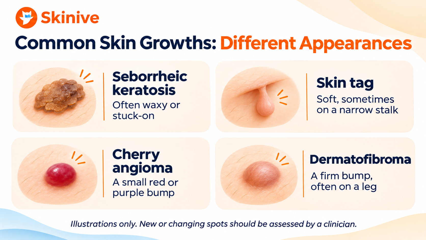
| Type | How it may look or feel | Common locations |
|---|---|---|
| Seborrheic keratosis | Waxy or rough growth with a “stuck-on” appearance | Face, neck, chest or back |
| Skin tag | Small, soft growth, sometimes on a narrow stalk | Neck, armpits, eyelids or groin |
| Cherry angioma | Small red or purple spot or bump | Often the trunk |
| Dermatofibroma | Firm, rounded skin bump | Often the legs |
| Solar lentigo | Flat brown pigmented patch | Sun-exposed skin, including face and hands |
| Sebaceous hyperplasia | Small skin-colored or yellowish bump, sometimes with a central indentation | Forehead or central face |
| Epidermoid cyst | Rounded lump beneath the skin, sometimes with a central opening | Various body areas |
| Milia | Tiny white or yellowish bumps | Face, especially around the eyes and cheeks |
| Lipoma | Soft lump beneath the skin that may move slightly | Shoulders, back, arms or thighs |
These descriptions are general. A lesion does not need to match every feature, and similar-looking lesions can have different causes.
When Should You See a Doctor About a Skin Lesion?
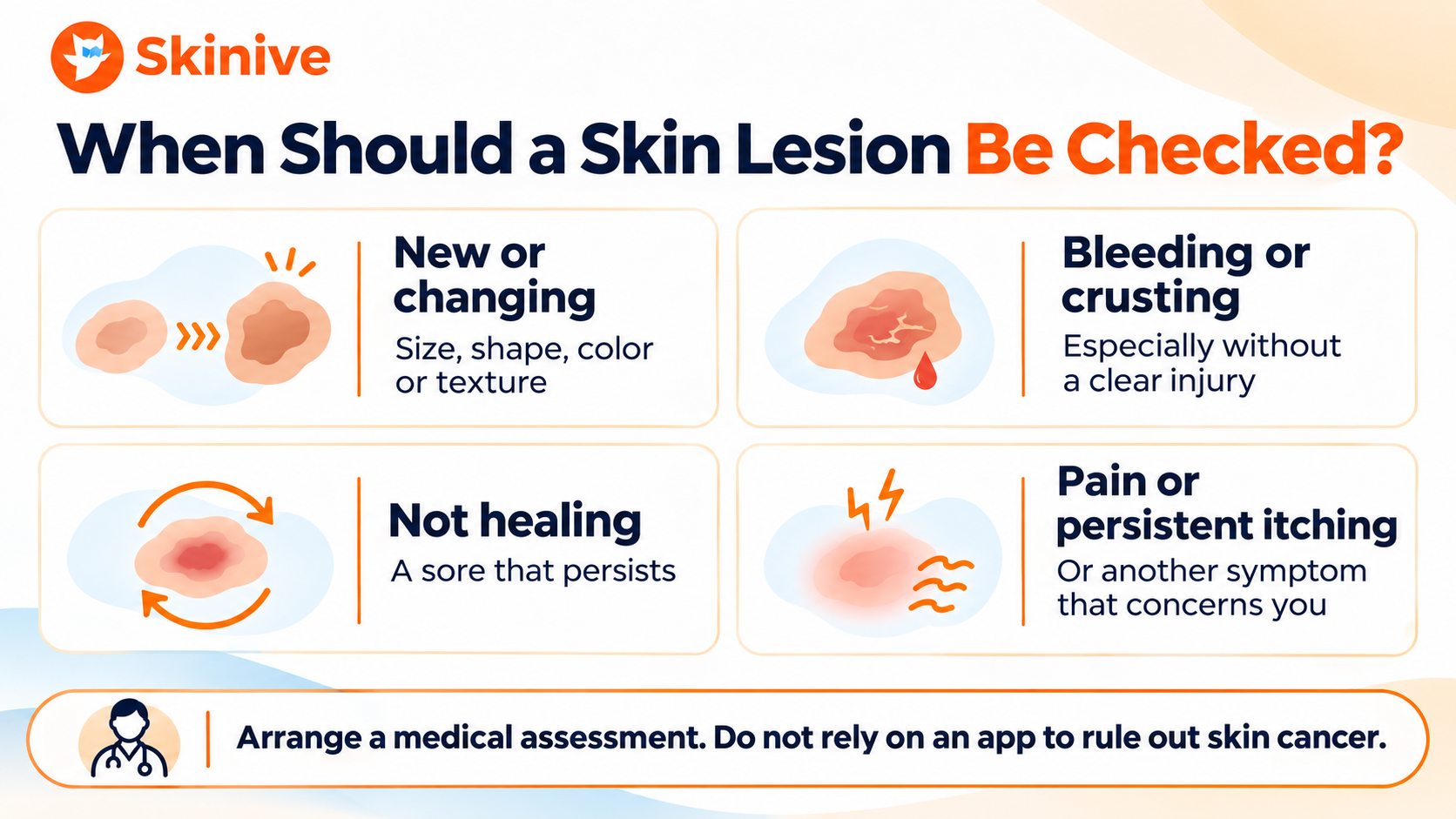
Arrange a medical assessment if you notice a skin lesion that:
- Is new or changing
- Changes in size, shape, color or texture
- Looks noticeably different from your other spots
- Bleeds, crusts, oozes or forms a sore that does not heal
- Causes persistent itching, tenderness or pain
- Grows quickly or develops into a lump that is hard or difficult to move
- Causes concern, even if you are unsure how to describe it
A concerning skin change can be small, painless or difficult to notice. Do not wait for several warning signs to appear.
Seek prompt medical advice for a rapidly worsening, painful, hot or spreading skin change, particularly if you also feel unwell or have a fever.
If you need help finding professional care, explore Skinive’s dermatologist resources.
Can a Benign-Looking Skin Lesion Be Skin Cancer?
Some skin cancers can resemble benign skin growths. Conversely, a harmless lesion may look unusual. A healthcare professional may use a skin examination, dermoscopy or, when needed, a biopsy to determine the cause.
For pigmented spots and moles, the ABCDE rule can help you notice asymmetry, irregular borders, uneven color, diameter and evolution. However, skin cancer does not always follow this pattern. A melanoma can also be smaller than 6 mm.
Explore melanoma in the Skinive Dermatology Atlas and learn more in our guide to types of skin moles and when to get them checked.
How to Monitor a Skin Lesion
Knowing what is normal for your skin makes it easier to notice a change. A regular skin self-check can help you become familiar with your moles, spots and other visible skin features.
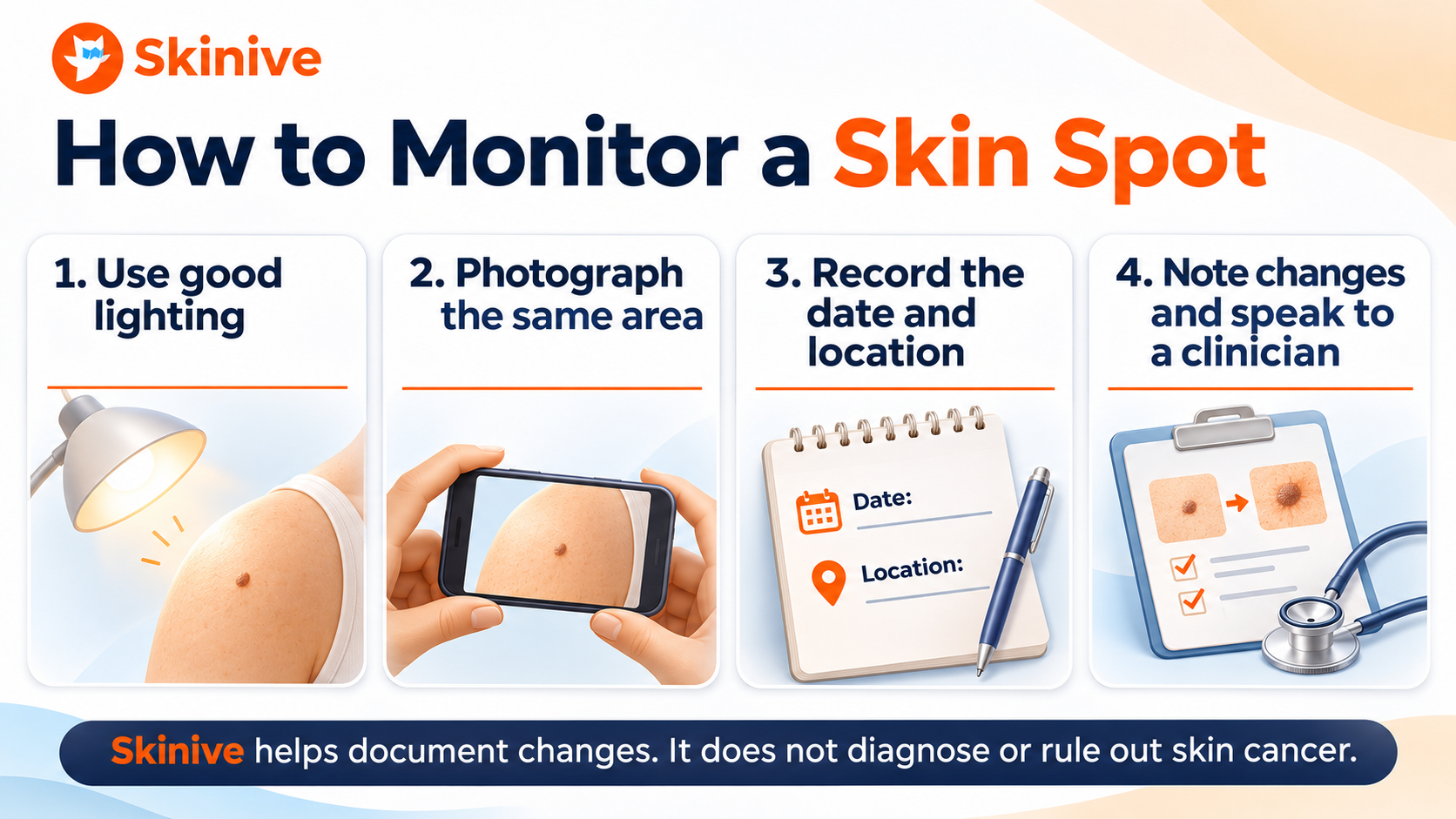
When documenting a spot:
- Take a clear photo in good lighting.
- Record the date and the location on your body.
- When taking follow-up photos, use a similar angle, distance and lighting where possible.
- Note changes in size, shape, color, texture or symptoms.
- Share your observations with a healthcare professional if anything changes or concerns you.
A photograph is a record, not a diagnosis. If a spot already looks concerning, seek medical advice rather than waiting to see whether it changes further.
Can Skinive Help You Monitor Skin Changes?
Skinive helps users document visible skin concerns with photographs and review previous checks over time. Depending on the feature, you can explore information about moles, acne, pigmentation and other visible changes.
You may find these Skinive tools useful:
- AI skin checker and mole tracker: Learn about documenting and monitoring visible skin concerns.
- Skin cancer screening app information: Understand how image-based tools can support skin awareness and where their limits are.
- Acne Tracker: Follow visible acne-related changes over time.
- Face Analysis app: Explore tools for observing visible facial skin concerns.
- UV Index app: Check UV conditions and forecasts for your location.
- Sun Exposure and Tanning Timer: Follow time outdoors alongside information such as the UV Index, selected skin type and SPF.
Skinive does not diagnose or rule out skin cancer or other medical conditions. Image-based results may be incorrect or inconclusive. A new, changing, bleeding or otherwise concerning lesion should be assessed by a qualified healthcare professional, regardless of an app result.
Can You Prevent Skin Lesions?
Not every skin lesion can be prevented. Some are associated with age, genetics or factors that are not fully understood.
You can, however, take practical steps to protect your skin:
- Use shade and protective clothing when UV exposure is significant.
- Apply broad-spectrum sunscreen with SPF 30 or higher to exposed skin and reapply as directed.
- Avoid indoor tanning.
- Check your skin regularly and seek advice about concerning changes.
- Avoid picking, squeezing or attempting to remove skin growths yourself.
The UV Index can help you understand current outdoor conditions. Exposure-time estimates are approximate and should not be treated as a guarantee against sunburn or skin damage.
Read more about choosing and applying sunscreen.
Frequently Asked Questions
Are all skin lesions dangerous?
No. Many skin lesions are benign. However, a new or changing spot cannot be identified with certainty from a general description alone. If you are concerned, consult a healthcare professional.
What is the difference between a skin lesion and a mole?
A skin lesion is a broad term for an area of skin that looks or feels different. A mole is one type of skin lesion. Other examples include skin tags, cysts and sunspots.
Can a skin tag turn into skin cancer?
A confirmed skin tag is benign. However, other growths can resemble skin tags. Have a new, changing, bleeding or uncertain growth examined before considering removal.
Do all skin lesions need to be removed?
No. Many confirmed benign lesions need no treatment. Removal may be considered if a lesion is irritated, symptomatic, cosmetically bothersome or needs further examination.
Can I identify a skin lesion from a photo?
A photo can help you document a skin change or learn about possible appearances, but it cannot establish a reliable diagnosis on its own. A clinician can consider your symptoms, history and examination findings.
When should I have a lump under my skin checked?
Have a new lump assessed by a healthcare professional. A lump that grows, becomes painful, feels hard or does not move should be brought to a clinician’s attention.
Can an app tell me whether a skin lesion is cancerous?
No. An app can support documentation and skin awareness, but it cannot confirm or rule out cancer. Seek professional advice for a new, changing or otherwise concerning lesion.


